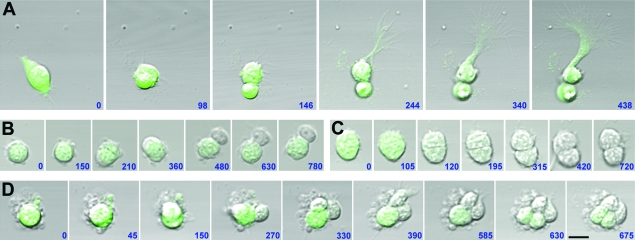
Figure 2.
Analysis of isolated cortical progenitor development. Radial progenitors micro dissected from the VZ of E14.5 BLBP-GFP embryonic cortices were plated at clonal density and isolated, and genetically defined radial progenitors (GFP+) were time-lapse imaged. (A) Division of GFP+ progenitor resulting in 2 other GFP+ cells, 1 of which extends a basal process. (B) Division of GFP+ progenitor resulting in 1 GFP+ cell and 1 GFP− cell. (C) Division of GFP+ progenitor resulting in 2 GFP− cells. (D) Generation of a mixed clone containing GFP+ and GFP− cells. The combined use of time-lapse analysis of genetically defined cortical progenitors with correlative immunolabeling of daughter cells at the end of imaging can be used to study patterns of normal and altered progenitor development. Time elapsed between observations are indicated in minutes. Simultaneous GFP and phase light images were obtained using a Zeiss inverted confocal microscope equipped with live-cell incubation chamber. Scale bar = 25 μm.

