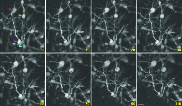
Figure 3.
In utero imaging of interneuronal dynamics. GFP+ interneurons in the developing cerebral cortex (parietal cortical area) of E16.5 Dlx5/6-CIE embryos were repeatedly imaged using 2-photon microscopy. Time-lapse imaging of these neurons in living embryos illustrates dynamic changes in their position, process growth, or neighbor relationships. Arrowheads indicate sample neurons undergoing such changes. Time elapsed is indicated in minutes. Scale bar = 30 μm.

