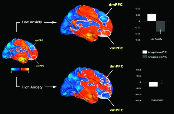
Figure 3.
Resting-state functional connectivity of the amygdala with the rest of the brain, divided into high (N = 15) and low (N = 14) anxiety groups. The low anxiety group is characterized by a strong positive amygdala–vmPFC connectivity and negative amygdala–dmPFC connectivity. In contrast, the high anxiety group showed negative amygdala–vmPFC connectivity and disrupted amygdala–dmPFC connectivity. The white ovals depict approximate locations of the voxel clusters shown in Figure 2. Error bars indicate the standard error of the mean.

