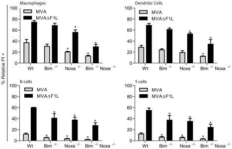
Figure 4. Contributions of Noxa and Bim to apoptosis induced by MVA and MVAΔF1L in primary MVA target cells.
M-CSF bone marrow derived macrophages and GM-CSF bone marrow derived dendritic cells, B220+ MACS sorted B-cells and NK1.1−/B220−/MHCII− MACS sorted ConA activated T-cells were infected with MVA or MVAΔF1L at an M.O.I of 10 for 20 h. Cell death was assessed by PI staining and relative numbers were calculated by calculating the % increase in PI proportional to the untreated sample with the following equation: (((% treated PI+)−(% untreated PI+))/% untreated PI+*100). (* indicates statistical significance according to the student's t-test, p≤0.05; with data giving mean/SEM of n≥3).

