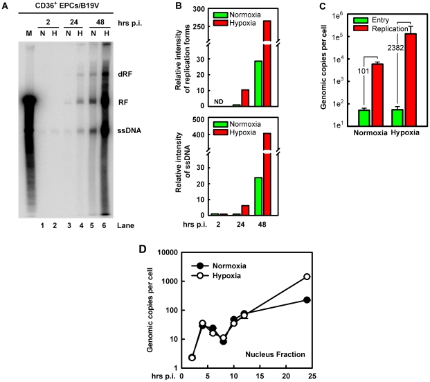
Figure 3. B19V DNA replication, virus entry and intracellular trafficking in CD36+ EPCs under hypoxia vs. normoxia.
B19V DNA replication, entry and trafficking were assessed in day 8 CD36+ EPCs infected with B19V at an MOI of 5,000 gc/cell under each condition. (A&B) At the indicated times p.i., Hirt DNA was extracted and analyzed by Southern blotting. (A) The full blot is shown. M, marker, a 5.6-kb B19V DNA; N, normoxia; H, hypoxia; RF, replicative form DNA; dRF: double replicative form DNA; ssDNA: single-stranded DNA. (B) The bands of the viral RF DNA and ssDNA genome in the blot were quantified and values shown are relative to the normoxia-cultured sample of either collected at 24 hrs p.i. (upper panel) or at 2 hrs p.i. (lower panel). (C) B19V entry into the cell and virus replication were assessed. The entered number of viral genomes within the cell under each condition was assessed, and is shown in green bars. Duplicated sets of treated cells were maintained under the respective condition until 48 hrs p.i., at which point total viral DNA was quantified. Results are shown as absolute number of genomic copies per cell in red bars. (D) The rate of B19V trafficking to the nucleus and replication was measured in infected cells, at the indicated time points. The numbers of genome copies per cell in the nucleus are shown.

