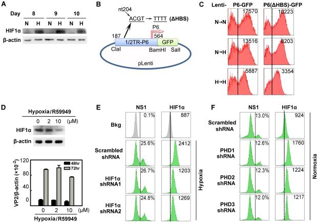
Figure 5. Regulation of B19V infection of CD36+ EPCs by HIFα under hypoxia vs. normoxia.
(A) HIF1α levels in CD36+ EPCs cultured under normoxia (N) or hypoxia (H). Cell lysates were prepared on the indicated days and were analyzed by Western blotting with anti-HIF1α and anti-β-actin. (B&C) Effects of the putative HIF-HBS in the P6 promoter. Day 7 hypoxia- or normoxia-cultured CD36+ EPCs were transduced with Lenti-P6-GFP or Lenti-P6(ΔHBS)-GFP (B), and then were either maintained under normoxia (→N) or hypoxia (→H). At 48 hrs post-transduction, level of GFP expression (as MFI) was measured by flow cytometry. (D) Effects of R59949 on B19V infection. Day 8 hypoxia-cultured CD36+ EPCs were treated with R59949 at the final concentrations shown. At 24 hrs post-treatment, cells were collected for HIF1α detection by Western blotting, or infected with B19V at an MOI of 1,000 gc/cell. At 48 and 72 hrs p.i., the levels of VP2-encoding mRNA per β-actin mRNA were quantified. (E&F) Effects of HIF1α and PHD knockdown on B19V infection. Day 7 CD36+ EPCs cultured under each condition were transduced with the indicated lentiviruses. At 48 hrs post-transduction, cells were infected with B19V (at MOIs of 2,000 and 4,000 gc/cell for hypoxia- and normoxia-cultured cells, respectively). At 48 hrs p.i., the expression levels of NS1 (as % of positive cells) and HIF1α (as MFI) were analyzed by flow cytometry in lentivirus-transduced (GFP+) cells. Dashed reference lines were selected arbitrarily to show the relative position of the peaks, and Bkg (background) represents the secondary antibody only control.

