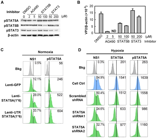
Figure 7. Regulation of B19V infection of CD36+ EPCs by phosphorylated STAT5A.
(A&B) Effect of inhibiting STAT phosphorylation on B19V infection. Day 8 CD36+ EPCs cultured under normoxia were pre-treated with pharmacological inhibitors of phosphorylation at the indicated final concentration, or with 0.5% DMSO (control). At 24 hrs post-treatment, the cells were analyzed by Western blotting for the inhibitory effects on phosphorylation (A); or were infected with B19V (at an MOI of 5,000 gc/cell) (B). At 48 hrs, p.i., the infected cells were assessed for the level of B19V VP2-encoding mRNA per β-actin mRNA. (C&D) B19V infection in the context of constitutively active STAT5A and STAT5A-specific shRNAs. Day 7 CD36+ EPCs cultured either under normoxia (C) or hypoxia (D) were transduced with the indicated lentivirus. At 48 hrs post-transduction, the cells were infected with B19V at an MOI of 5,000 gc/cell, and at 48 hrs p.i., they were analyzed by flow cytometry for the expression of B19V NS1, STAT5A and pSTAT5A. The GFP-positive population of lentivirus-transduced cells was selectively gated. Percentages shown in the left column indicate the level of NS1-expressing cells, and the numbers in the right column the MFI of STAT5A or pSTAT5A. Results shown are representative of at least two independent experiments. Dashed reference lines in panels C&D were selected arbitrarily to show the relative positions of the peaks, and Bkg (background) represents the secondary antibody only control.

