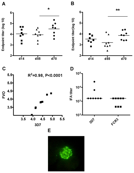
Figure 2. P. falciparum AMA1 specific total IgG responses induced by immunization in rabbits.
New Zealand rabbits (n = 8) were immunized via the intradermal route with 5×10¹⁰ vp of AdHu5-PfAMA1 (3D7) and boosted 8 weeks later with 5×10⁷ pfu of MVA-PfAMA1 (3D7). The AMA1-specific antibody response against both homologous 3D7 (A) and heterologous FVO (B) PfAMA1 recombinant protein was measured in the serum at day 14 (post prime), 55 (pre boost) and 70 (post boost). Data show the responses from individual rabbits and the lines represent the median. The asterisk indicates a significant difference between the different time-points as analyzed by Wilcoxon signed rank test (* P ≤0.05, ** P ≤0.01). (C) The correlation between the 3D7 and FVO antibodies at day 70 was analysed using Pearson's rank correlation. There was a highly significant positive correlation (R2 = 0.96, P ≤0.0001). (D) IFA titers were measured using sera taken two weeks following the boost immunization. Individual IFA titers against 3D7 and FCR3 P. falciparum strains are shown with the median response (FCR3 AMA1 differs from FVO AMA1 by only a single amino acid substitution [18]). (E) When serum from immunized rabbits was used to label P. falciparum schizonts, typical AMA1-specific punctate apical staining of merozoites was seen.

