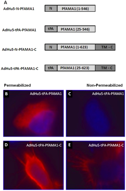
Figure 4. Schematic representation of the different PfAMA1 (3D7) constructs and effect of the addition of the C terminus on cellular localization of PfAMA1.
(A) The figure shows the different AdHu5 vaccine inserts generated to test the effect of replacing the human tPA leader sequence with the native AMA1 N-terminal predicted signal peptide (aa 1–24) and also the effect of addition of the native transmembrane domain and C-terminal cytoplasmic domain (aa 547–623). PfAMA1 in this figure represents the ectodomain of P. falciparum 3D7 AMA1 (aa 25–546). Hela cells were grown on cover slips and transfected with the plasmid encoding tPA-PfAMA1 (B and C) or tPA-PfAMA1-C (D and E). After overnight incubation polyclonal serum from immunized mice was used to detect the localization of PfAMA1. Alexa-conjugated anti-mouse antibody was used as the secondary antibody and the DNA was stained with 4′, 6-diamidino-2-phenylindole (DAPI). Cell surface localization of PfAMA1, notably on the cell projections, was seen in the construct that has the C terminal domains in both the permeabilized (D) and non-permeabilized samples (E). There is no cell surface associated PfAMA1 in the construct without the C terminal domains (B and C).

