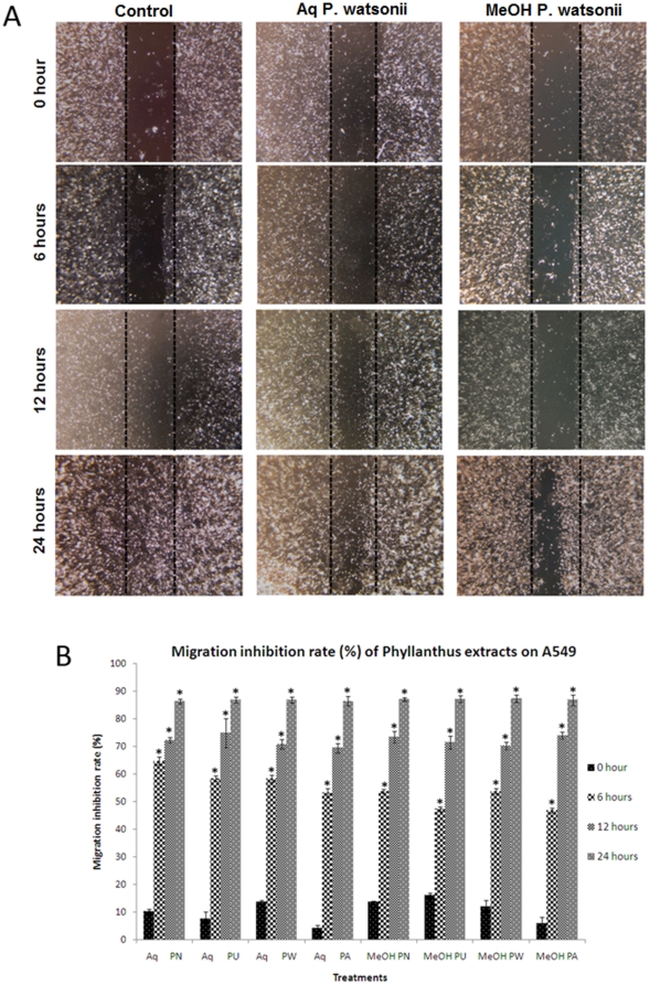
Figure 2. Wound closure activity of treated A549 cells after 24 hours.
(A) Representative photographs of wounded A549 cell monolayer treated with aqueous and methanolic P. watsonii extracts. Typical result from three independent experiments is shown. (Magnification power: 200×) (B) Quantitative assessment of migration inhibition rate of Phyllanthus on A549 cells. Error bar indicates the standard error of the mean of three independent experiments. PN – P. niruri, PU – P. urinaria, PW – P. watsonii, PA – P. amarus. *P<0.05 vs control.

