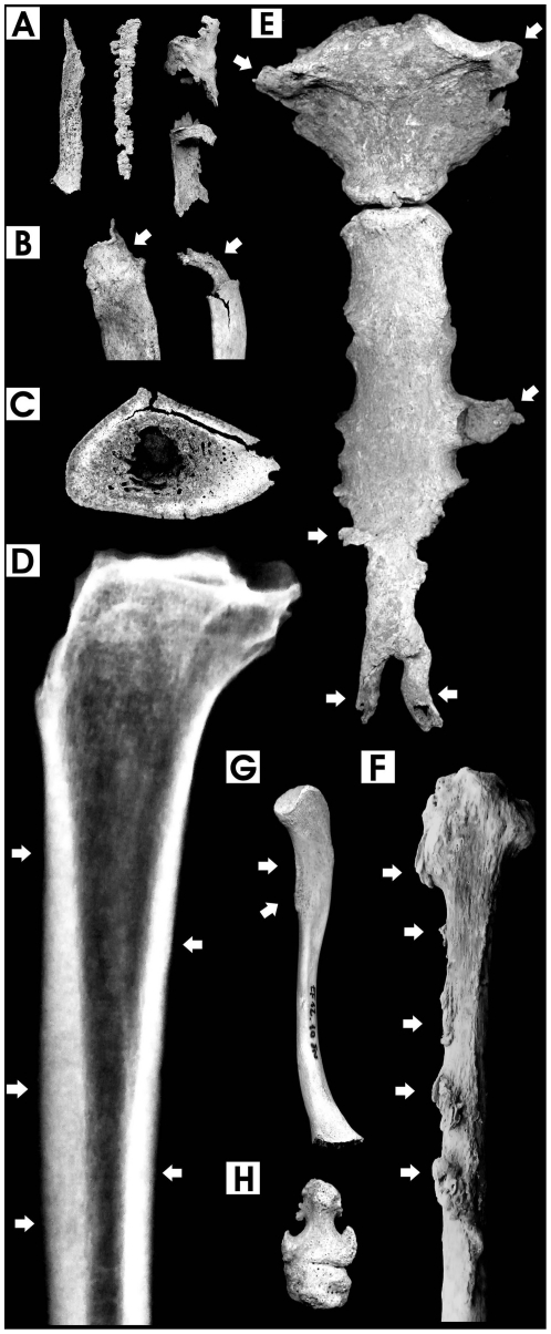
Figure 1. Pathological features in chest and long bones.
A. Calcified ligaments and interosseous costal cartilages, 40-year-old male; B. Proximal first and sixth rib epiphyses with prominent exostoses due to interosseous cartilage calcification, 27-year-old female and 40-year-old male; C. Cross section of the mid-shaft of tibia showing extensive cortical thickening, increased bone matrix density, intracortical resorption and reduced medullary space, 40-year-old male; D. Digital x-ray image (lateral view) of the previous tibia, showing a “marble-like” appearance (arrows) symptomatic of marked osteosclerosis; E. Prominent calcification of costosternal and costoxiphoid ligament attachments (arrows) in the sternum, 40-year-old male; F. Ligamentous and interosseous membrane ossification at multiple sites (arrows) in the fibula, 40-year-old male; G. Calcification and osteophytosis at the attachment of the deltoid muscle (arrows) in the clavicle, 9-year-old male; H. Ankylosis of toe distal interphalangeal joint, 29-year-old male (bone images are in 1∶2 size).

