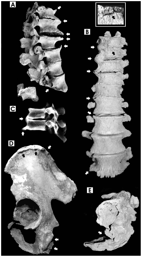
Figure 2. Pathological features of spine and pelvis.
A. Widespread hypertrophic osteosclerosis, calcification of anterior ligaments, spondyloarthrosis and osteoporosis (arrows) of thoraco-lumbar vertebrae (T12-L5, lateral view), 44-year-old male. Notice severe flattening (osteoporosis) of the L5 vertebral body (arrow) and lumbar spondylolysis (inferior articular part split separately from the spinous process); B. Healed fracture of T10 (see enlargement in the small box), severe calcification of thoraco-lumbar anterior ligaments (T9-L5, anterior view) and ankylosis of T9-T11 vertebrae (arrows), 52-year-old male. Spondylolysis affects the L5 vertebra too; C. Digital X-ray image of T8-T9 fused vertebrae, showing diffuse osteosclerosis (lateral view), 38-year-old male; D. Ligamentous calcification and osteophytic bony spurs at the iliac crest and ischial tuberosity (sacrotuberous ligament) (arrows), 52-year-old male; E. Healed fracture of the 3rd vertebra (arrow) and kyphosis of the sacrum bone (lateral view), 52-year-old male (bone images are in 1∶2 size).

