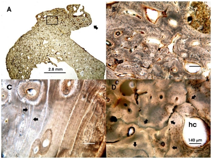
Figure 3. Histopathological bone features.
Representative bone ground sections observed under transmitted light microscope: A. Mid shaft of fibula showing increased cortical thickness, reduced medullary cavity and a prominent exostosis (arrow) abnormally exceeding the original shape of the bone surface, 40-year-old male; B. Higher magnification of the insert of figure A, showing bone cancellization, enlarged Haversian systems and disordered lamellar architecture; C. Mid shaft of femur with widespread deficiency of the Haversian lamellar systems (arrow), extensive mottling of bone matrix and several enlarged Haversian canals, 15-year-old male. Note the presence of several linear formation defects (arrows); D. Mid shaft of tibia with osteonic texture locally poor, mottled bone matrix and an extremely enlarged Haversian canal (hc), 30-year-old female. Note the extensive and irregular cracking (arrows) induced by exposure of victims' corpses to the hot pyroclastic surge. B, C and D images are at the same magnification.

