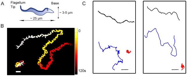
Figure 1. Trypanosome trajectories.
a) Scheme of a typical trypanosome cell shape with dimensions. The flagellum originates at the flagellar pocket, near the kinetoplast and runs along the entire body b) Time-lapse overlay of trypanosomes trajectory illustrates tumbling (lower left), and running motion (upper right), scale bar  , colorbar represents time, both trajectories represent about 2 minutes of trypanosome swimming. c) Diversity in trypanosome trajectories, reveals three motility modes in which cells tumble (tumbler, in red), travel directionally (persistent, in black), or alternate between tumbling and running motion (intermediate, in blue). Left panel derived from experiment, right panel from model, scale bars
  .
.

