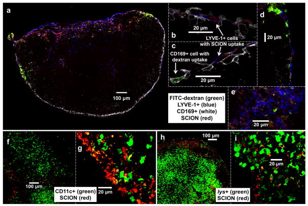
Fig. 7.
Confocal microscopy of SCION(AlexaFluor555) localization in politeal lymph node 24 hr post-intradermal injection in hind footpad. Panels b-e, g, and i are magnified regions of interest from LNs panels a, f, and h which capture the LNs as a whole or from their circumference. In all nodes, SCION was primarily found in the subcapsular space, trabelculae, and near the exiting efferent vessel in the medullary and hilum regions. (a-e) B6-albino mouse co-injected with FITC-dextran that localized in CD169+ subcapsular macrophages, whereas SCION particle was observed in LYVE-1+ endothelial cells lining the subcapsular floor (b-d) and hilum (a,e). (f-g) CD11c-EYFP mouse showed minimal uptake of SCION by CD11c+ dendritic cells. (h-i) lys-EGFP mouse biopsy found minimal overlap between SCION and lys+ myelomonocytic cells (neutrophil granulocytes and macrophages)

