
Figure 2.
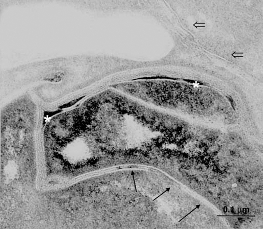
Abnormal SC membranes in SLS skin. Note the paucity of membrane bilayers in some regions (arrows) and lamellar domains that are interspersed with lacunae filled with non-lamellar lipid material (asterisks). Entombed lamellar contents in can be seen in corneocyte cytosol (open arrows). Ruthenium tetroxide post-fixation. Reprinted from Rizzo et al. Arch Dermatol Res 2010; 302:443.