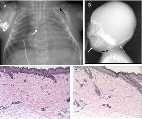
Figure 2. Radiographs and histopathology of skin biopsy of RD 500.3.
A. Anterior-posterior chest radiograph of patient RD 500.3 shows a butterfly vertebra at the thoracic 5 level (white arrow). Clavicles were dysmorphic with a pseudoarthrosis at the juncture of the middle and distal one-thirds with the distal portions bulbous in appearance (black arrow).
B. Lateral radiograph of the skull of patient RD 500.3 reveals prominent sutures (gray arrow), micrognathia (white arrow), and hypoplastic cervical vertebral bodies (black arrow).
C. Hematoxylin and eosin stain of skin biopsy shows the epidermis and dermis to be slightly attenuated, and the dermis shows marked fibrosis and parallel collagen bundles.
D. An elastic tissue stain, Verhoeff van Gieson, shows complete absence of elastic fibers throughout the dermis.

