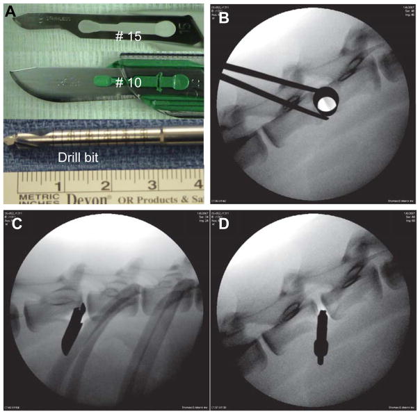
Figure 1. Instruments used to induce disc injury in the goat and sample X-rays obtained during surgery.
A. The instruments used to create disc injuries (from top to bottom: #10 blade, #15 blade, 4.5 mm drill bit); B–D: lateral X-ray of the goat spine documenting the positions of the instruments: B. large dilator inserted adjacent to the intervertebral disc; C. the #10 blade; D. the 4.5 mm drill bit.

