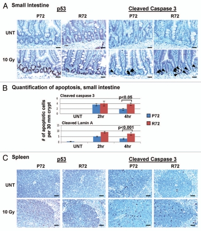
Figure 2.
Tissue-specific effects of the codon 72 polymorphism on apoptosis. (A) Immunohistochemistry using antisera for p53 and cleaved caspase 3 was performed on the small intestine of P72 and R72 mice that were untreated (UNT) or 4 h after 10 Gy gamma radiation. Increased levels of cleaved caspase 3 staining are visible in the crypt of R72 mice 4 hours after gamma radiation while the levels of p53 staining are comparable between P72 and R72 mice. The data depicted are representative of five independent experiments. Arrowheads: apoptotic cells. Scale bar: 50 um. (B) Quantification of cells positive for cleaved caspase 3 and cleaved lamin A in the small intestine of P72 and R72 mice 4 h after 10 Gy. The number of positive cells per 30 mm of crypt was counted and tabulated; the results shown are averaged from three independent experiments, and standard deviation bars are shown. p values were calculated using the Student's two-tailed t-test. Scale bar: 50 um. (C) Immunohistochemistry using antisera for p53 and cleaved caspase 3 was performed on the spleens of untreated (UNT) P72 and R72 mice or 4 hours after 10 Gy gamma radiation. The data depicted are representative of five independent experiments.

