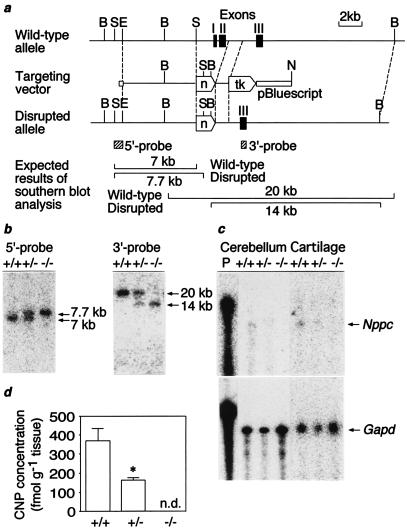
Figure 1.
Targeted disruption of the mouse Nppc. (a) Restriction maps of the wild-type 129/Sv mouse Nppc allele, targeting vector, and the predicted disrupted allele. Closed boxes indicate exons (I–III). Locations of 5′ and 3′ external probes are shown as hatched bars. B, BamHI; S, SphI; E, EcoRI; N, NotI; tk, Herpes simplex virus thymidine kinase gene; n, neomycin resistance gene. (b) Southern blot analysis of genomic DNAs from F2 offspring with 5′ and 3′ probes upon digestion with SphI and BamHI, respectively. (c) RNase protection analysis of Nppc and Gapd transcript in the cerebellum and tibial epiphyseal cartilage. The cerebellum and tibial cartilage are obtained from mice at 20 weeks and 7 days of age, respectively. Ten micrograms and 1 μg of total RNA were used to analyze Nppc and Gapd transcripts, respectively. (d) Cerebellar CNP concentrations at 20 weeks of age (n = 4). n.d., not detectable. *, P < 0.05 vs. Nppc+/+ mice.

