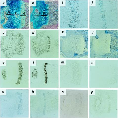
Figure 3.
Histological analysis of proximal epiphyseal cartilages of the tibia from 7-day-old Nppc+/+ and Nppc−/− mice. (a and b) Alcian blue-hematoxylin/eosin staining of tibial epiphyseal cartilages from Nppc+/+ (a) and Nppc−/− (b) mice. Resting (R), proliferating (P), and hypertrophic (H) zones are indicated. (c–j, m–p) In situ hybridization analysis of tibial epiphyseal cartilages from Nppc+/+ (c, e, g, i, m, o) and Nppc−/− (d, f, h, j, n, p) mice showing expression of mRNA for Col2a1 (c, d), Col10a1 (e, f), Ihh (g-j), Nppc (m, n), or Npr2 (o, p). (k and l) Immunohistochemical detection of BrdUrd-labeled chondrocytes in the tibial growth plate from Nppc+/+ (k) and Nppc−/− (l) mice. (Magnification: a–h, k–p, ×40; i, j, ×100.)

