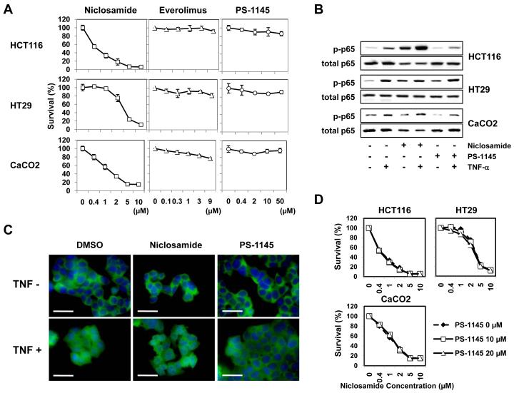
Figure 3. The mTOR and NF-κB pathway were not targets of niclosamide.
(A) Colorectal cancer cell lines (HCT116, HT29, CaCO2) were treated with niclosamide (0-10 μM), everolimus (0-9 μM), or PS-1145 (0-50 μM) for 72 h, and cytotoxicity was analyzed in MTT assays. The relative ratio (percentages) of OD 562 nm values were calculated for each reagent by setting the OD 562 nm value at concentration 0 μM as 100%. (B, C) To examine if the NF-κB pathway could be perturbed by niclosamide, cancer cells were treated with niclosamide (1 μM) or PS-1145 (10 μM) for 24 h, and further incubated with TNF-α (10 ng/mL) for 1 h. Whole cell lysates were made and analyzed with anti-phospho-NF-κB p65 and anti-p65 antibodies by Western Blot (B). Cells were fixed, permeabilized and stained with FITC-conjugated anti-NF-κB p65 antibody (green) for 1 h. Nuclei were stained with DAPI (blue). Staining of HCT116 cells is shown. Unclear morphology of nuclei (blue) indicates nuclear translocation of p65 protein (C). Scale bars: 30 μm. (D) To determine whether niclosamide-induced cytotoxicity of colorectal cancers could be prevented by blocking the NF-κB pathway, cancer cells were treated with niclosamide (0-10 μM) in the presence/absence of PS-1145 (10, 20 μM) for 72 h and an MTT assay was performed.

