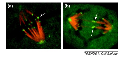
Figure II.
Merotelically attached kinetochores in PtK1 cells. PtK1 cells are ideal for studying kinetochores because of the small number of chromosomes and the fact that the cells remain flat throughout mitosis, making them amenable for high-resolution light microscopy. The images show live PtK1 cells microinjected with X-rhodamine-labeled tubulin and Alexa 488-labeled CENP-F antibodies to fluorescently label kinetochore fibers (red) and kinetochores/spindle poles (green), respectively. (a) Late prometaphase PtK1 cell with one merotelic kinetochore (arrow) aligned at the metaphase plate. (b) Anaphase PtK1 cell with two merotelically attached lagging chromosomes (arrows).

