
Figure 3.
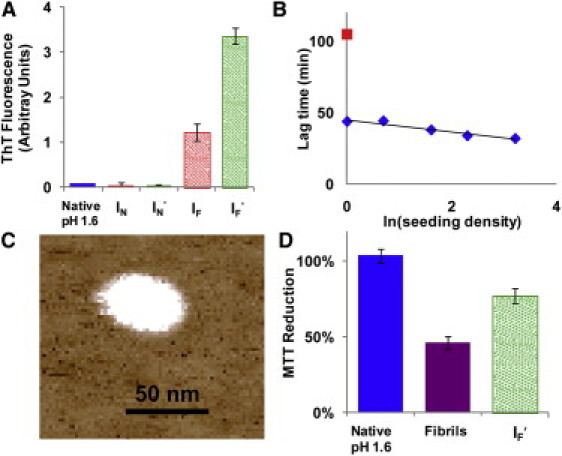
Amyloid characteristics of disaggregated insulin. (A) Thioflavin-T (ThT) fluorescence of insulin samples shows that disaggregated insulin (IF and IF′) has a ThT signal, whereas native insulin (IN and IN′) does not. (B) Seeding of IF′ into a new kinetic run reduces the lag time in a dose-dependent fashion (blue diamonds). For reference, the control without seeding is shown as a red square. (C) AFM of IF shows large aggregates 48 ± 15 nm in diameter at pH 11. (D) Fibrils and disaggregated fibrils (IF′) demonstrate cellular toxicity by an MTT reduction using PC-12 cells, whereas insulin that has not been in fibrils (Native insulin) does not reduce MTT. All MTT samples were added at pH 1.6, but the cell media buffered the samples to pH 7.4.