
Figure 1.
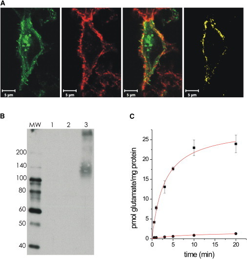
Heterologous protein expression and functional characterization of EAAT5. (A) AcGFP-EAAT5 expressing cells (green), counterstained with TRITC wheat germ agglutinin (red) exhibit 63% ± 6% membrane localization of EAAT5 (yellow, n = 5). (B) Immunoblot analysis reveals the typical broad electrophoretic mobility of EAATs (lane 1: nontransfected; lane 2: EAAC1-transfected; and lane 3: EAAT5-transfected cells). Molecular mass markers (lane MW) are indicated in kDa. (C) EAAT5 expression (solid squares) significantly increases L-[3H]-glutamate uptake in HEK293 cells (nontransfected HEK293, solid circles), which is in the same range as that measured for EAAT4 (19).