
Figure 1.
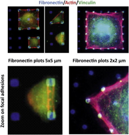
Fibroblasts adhering on fibronectin-patterned substrates. (Left panel) Cells adhering to 4 × 4 μm fibronectin plots. (Right panel) Cells adhering to 2 × 2 μm plots. Adhesive plots are visualized by Alexa350-labeled fibronectin. Actin filaments are labeled with TRITC phalloidin and focal adhesion by a monoclonal anti vinculin antibody and a Alexa488-labeled secondary antibody. For the largest plots, the focal adhesion complexes split into two parts with two stress fibers connecting opposite plots. For the smallest plots, however, the focal adhesive clusters appear homogeneous with only one stress fiber emerging from the adhesion complexes. The transition from one to two adhesive spots on the same plot is geometry-independent, as all cells adhering to square or triangular lattices exhibit the same behavior.