
Figure 1.
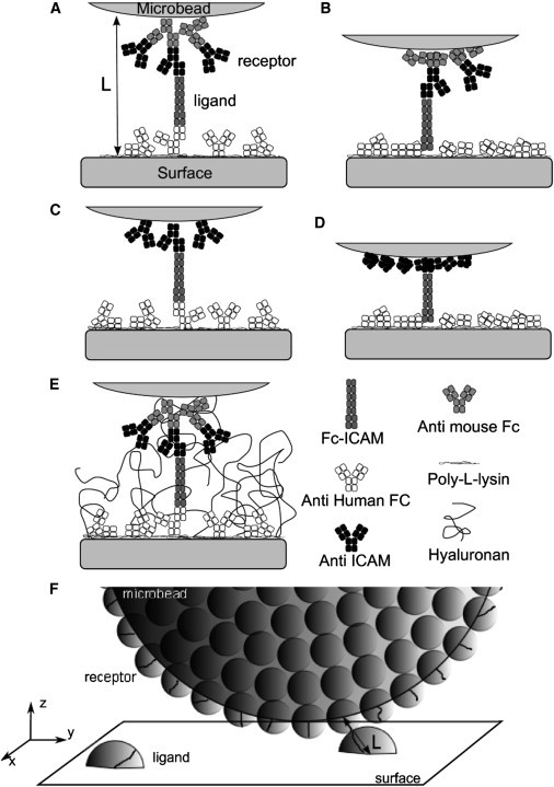
(A–E) Schematic representation of the different molecular constructions to probe ICAM-1 versus anti-ICAM-1 binding in the laminar flow chamber, at the onset of bond formation. Each square represents an Ig domain of 4 nm. (A and B) Configuration with a double layer (DL) of antibodies on the bead, with a maximal molecular tether length of LDL = 76 nm (A) or an intermediate extension tether length of 60 nm (B). (C and D) Configuration with a single layer (SL) of antibodies on the bead, with a maximal molecular tether length of LSL = 60 nm (C) or an intermediate extension tether length of 44 nm (D). (E) Configuration A in presence of adsorbed hyaluronan molecules acting as a repulsive layer. (F) Schematic representation of one microbead at the vicinity of the functionalized surface.