
Figure 3.
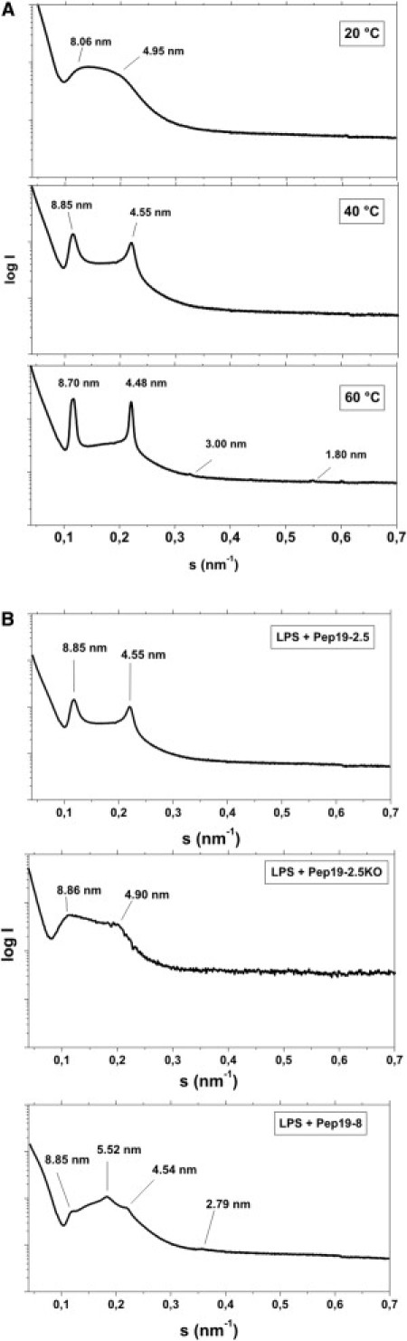
Supramolecular aggregate structure of LPS. SAXS was performed using synchrotron radiation of LPS R60 in the presence of Pep19-2.5 at (A) three temperatures and (B) at 40°C in the presence of Pep19-2.5 (top), Pep19-2.5KO (middle), or Pep19-8 (bottom). All samples were prepared at [LPS]/[Pep] 3:1 weight %. The logarithm of the scattering intensity logI is plotted versus the scattering vector s (= 1/d, d = lattice spacings).