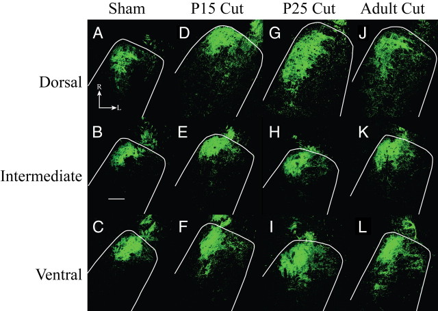
Figure 6.
Horizontal sections of labeled chorda tympani nerve terminal field. A–J, Fluorescent micrographs are shown for sham (A–C), P15 cut (D–F), P25 cut (G–I), and adult cut (J–L) rats. The location of the NTS is outlined in white. A, D, G, J, The dorsal zone of labeled chorda tympani nerve terminal fields expanded in the caudal and lateral directions in all groups compared with sham. B, E, H, K, There was little expansion of the chorda tympani nerve terminal field in the intermediate zone. C, F, I, L, Ventral zone sections showed increased chorda tympani nerve terminal field across all groups compared with sham. Scale bar, 200 μm. R, Rostral; L, lateral.

