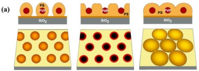
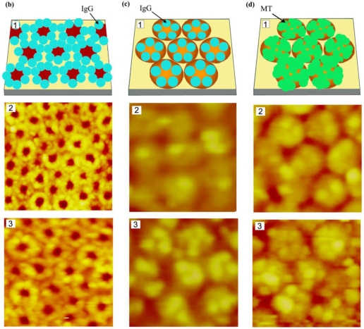
Figure 5.
Two-dimensional diblock copolymer templates of PS-b-PVP and protein assembly behavior observed on them; (a) various nanoscale templates resulting from chemical modification of nanodomains in micellar-forming diblock copolymers, (b and c) immunoglobulin G molecules on (b) open and (c) reverted PS-b-PVP templates, and (d) mushroom tyrosinase molecules assembled on a reverted PS-b-PVP template. The atomic force microscopy (AFM) scan size in panels (b) through (d) corresponds to (b): (2) 300 × 300 nm, (3) 180 × 180 nm, and (c and d): (2) 300 × 300 nm, (3) 180 × 180 nm. Adapted with permission from [100].


