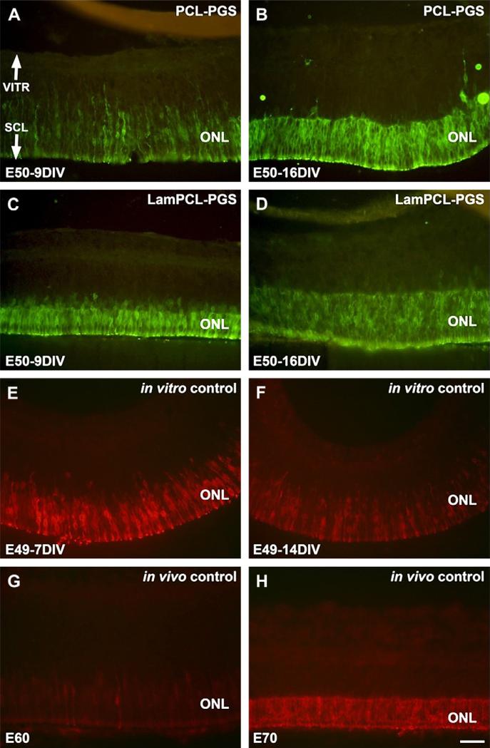
Fig. 5.
Rhodopsin staining of fetal porcine retina. A–D: E50 retinas co-cultured for 9 (A, C) and 16 (B, D) days in vitro with PGS membranes coated with PCL (A, B) or laminin-PCL blend (C, D) nanofibers. Rhodopsin labeled rod photoreceptors are visible in the outer nuclear layers of retinas, and survive at least 16 days in co-culture with PGS-nanofiber membranes. E–F: E49 retinas cultured for 7 (E) and 14 (F) days in vitro. Rhodopsin labeled rod photoreceptors are visible in the outer nuclear layer. G–H: E60 (G) and E70 (H) in vivo retina specimens. Rhodopsin labeled rod photoreceptors with neurites extending inwards as well as outwards to the margin of the retina are visible (G). Rhodopsin labeled rod photoreceptors are visible in the outer nuclear layer (H). VITR, vitreal (inner) aspect, SCL, sclera (outer) aspect. Scale bar = 50 microns.

