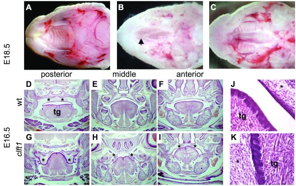
Figure 1. Clft1 palatal defects.
(A-C) E18.5 whole mount preparations with mandible and tongue removed. Clft1 mutants (B,C) show posterior cleft secondary palate, with normal elevation and fusion anteriorly (B), or complete cleft secondary palate (C). Arrowhead in B indicates closure of anterior palate. (D-K) Histological analysis at E16.5. Coronal sections from the posterior, middle and anterior portion of the oral cavity as indicated (*, palatal tissue; tg, tongue). (J,K) Higher magnification view of the oral adhesions between tongue and palate seen in clft1 mutants

