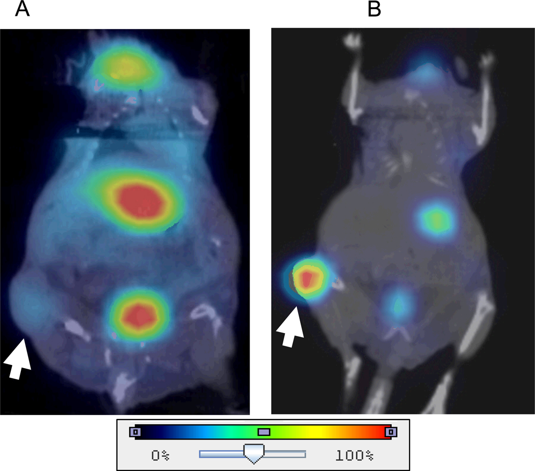
Fig. 1. Representative 123I micro-SPECT/CT images of BxPC-3 flank tumor-bearing nude mice.
A, Normal physiologic uptake only (arrow) (due to tumor vasculature and blood pooling in areas of tumor necrosis) was seen in the untreated control tumors. Also note areas of endogenous NIS-mediated radioiodide uptake in the thyroid, stomach, and accumulation in the bladder. B, In contrast, significant iodide accumulation (arrow) was seen in the MV-NIS infected xenografts.

