Figure 1.
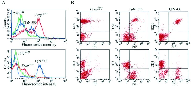
Analysis of PrPC expression on peripheral blood lymphocytes (PBLs) of TgN(Igκ Prnp)306 and TgN(CD19 Prnp)431 mice. (A) Single-stain FACS analysis for PrPC was as described in Materials and Methods; gating was for lymphocytes. PrPC expression on PBLs of TgN306 (Upper) and TgN431 mice (Lower) was compared with negative (Prnp0/0) and positive (Prnp+/+) controls. The two samples were analyzed on different occasions. (B) Double-color FACS analysis for PrP and B220 (Upper) and PrP and CD3 (Lower). PrP staining was followed by B lymphocyte staining with PE-conjugated anti-B220 antibodies or T lymphocyte staining with PE-conjugated anti-CD3e antibodies. PrPC expression was detected in B lymphocytes but not in T lymphocytes of both transgenic lines.

