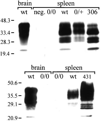
Figure 2.
Western blot analysis for PrPC in spleens of TgN(Igκ Prnp)306 and TgN(CD19 Prnp)431 mice. PrPC was enriched by immunoprecipitation with 6H4-linked Sepharose beads and was detected by using rabbit antiserum 1B3 against murine PrP (14). Crude wild-type and Prnp0/0 brain homogenates (120 μg total protein) were included in the experiment as controls for PrP immunodetection. Positions of the molecular weight standards (in kilodaltons) are indicated on the left side of the fluorogram. (Top) Expression of PrPC in the spleens of homozygous TgN306 mice. The equivalent of 10 mg of spleen was loaded per lane. A wild-type spleen sample immunoprecipitated with only Sepharose beads was included as a negative control (neg.). Genotypes of the mice analyzed: Prnp0/0,0/0; Prnp+/+, wt; Prnp0/+, 0/+; homozygous TgN306, 306. (Bottom) Expression of PrPC in the spleens of hemizygous TgN431 mice. Samples equivalent to 5 mg of spleen were loaded per lane. Genotypes of the mice analyzed: Prnp0/0, 0/0; Prnp+/+, wt; hemizygous TgN431, 431.

