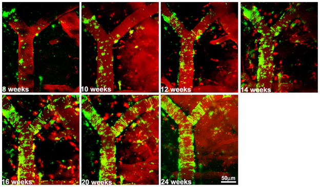
Fig. 2.
Age-dependent aggregation of CAA in a vessel segment. Repeated In vivo imaging of a PS1/APP mouse at two weeks intervals from 2 to 6-months of age. Serial imaging of the same volume in barrel cortex exhibited a progressive increase in cerebral amyloid angiopathy (labeled with methoxy-X04, green) in a typical vessel segment which was identified with rhodamine-dextran (red).

