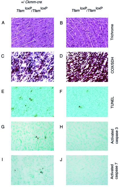
Figure 2.
Histology of hearts from Tfam heart knockouts (TfamloxP/TfamloxP, +/Ckmm-cre) and their littermate controls (TfamloxP/TfamloxP). Examples of immunoreactive cells are indicated by arrows. Trichrome stainings show no evidence for necrosis or fibrosis in Tfam knockout (A) or control (B) hearts. Double enzyme histochemical stainings for COX activity and succinate dehydrogenase activity show a mosaic loss of COX activity in Tfam knockout hearts, as evidenced by the blue staining of cardiomyocytes (C), and normal COX activity in controls, as reflected by the brown staining of cardiomyocytes (D). TUNEL stainings demonstrate more TUNEL-positive cardiomyocytes in Tfam knockout hearts (E) than in control hearts (F). Immunohistochemical stainings of cleaved caspase 3 and cleaved caspase 7 show occasional positive cardiomyocytes in Tfam knockout hearts (G and I) and no staining in control hearts (H and J).

