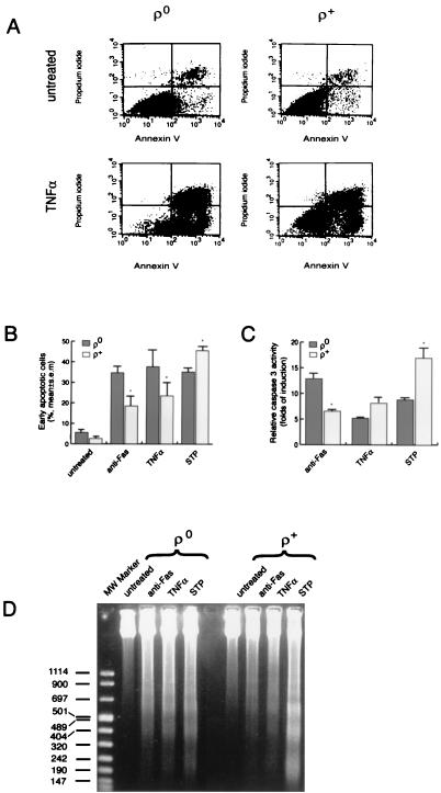
Figure 5.
ρ0 cells are susceptible to apoptosis induced by various signals. ρ0 (143B/206) and ρ+ (143B) osteosarcoma cells were incubated for 16 h with 0.5 μM STP, 100 ng/ml anti-Fas antibody plus 100 ng/ml actinomycin D (anti-Fas), or 20 ng/ml TNFα plus 100 ng/ml actinomycin D (TNFα). (A) Analysis by flow cytometry of apoptotic cells stained with annexin V and propidium iodide to distinguish early apoptotic cells (annexin positive, propidium iodide negative) from late apoptotic or necrotic cells (annexin positive, propidium iodide positive). (B) Susceptibility of ρ0 (143B/206) and ρ+ (143B) osteosarcoma cells to apoptosis in response to various signals as determined by annexin V/propidium iodide staining and flow cytometry. Values represent the percentage of early apoptotic (annexin V positive/propidium iodide negative) cells (%). *, P < 0.05. (C) Caspase 3 activities in ρ0 (143B/206) and ρ+ (143B) osteosarcoma cells. The results are plotted as fold induction of caspase 3 activity compared with untreated cells. *P < 0.05. (D) DNA ladders in ρ0 (143B/206) and ρ+ (143B) osteosarcoma cells.

