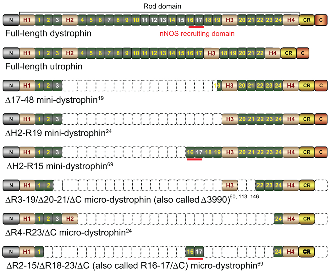
Figure 1.
Schematic illustration of full-length dystrophin, utrophin, and representative minidystrophin and microdystrophin.
Abbreviations: N, the N-terminal domain of the dystrophin protein; H1–4, hinges 1–4 in the rod domain of the dystrophin protein; numeric numbers, spectrin-like repeats in the dystrophin rod domain. Positively charged repeats are in white color. Repeats 11–17 represent the second actin-binding domain. CR, the cysteine-rich domain in the dystrophin protein; C, the C-terminal domain of the dystrophin protein; nNOS, neuronal nitric oxide synthase. Empty boxes denote the regions that are deleted in each respective minidystrophin and microdystrophin construct. Among three microgenes listed here, the ΔR3–19/Δ20–21/ΔC microdystrophin has been tested in affected dogs and human patients. However, these trials have failed to demonstrate a therapeutic efficacy. The hinge 2 region in ΔR4–R23/ΔC microdystrophin has been shown to compromise function. The potentially deleterious hinges 2 and 3 are removed in the ΔR2–15/ΔR18–23/ΔC microgene. Further, this microgene carries the nNOS localization domain.

