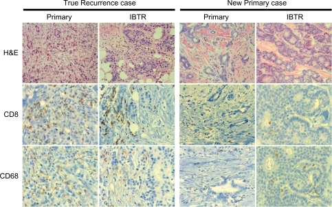
Figure 2.
Illustrative examples of two cases of primary tumors with the corresponding IBTR tumors. Cases were classified as TR IBTR (left) or NP IBTR (right). Panels show H&E, CD8, and CD68 immunohistochemical staining for primary and IBTR tumors in each case (original magnification 10× for H&E and 20× for CD3 and CD68).

