Figure 5.
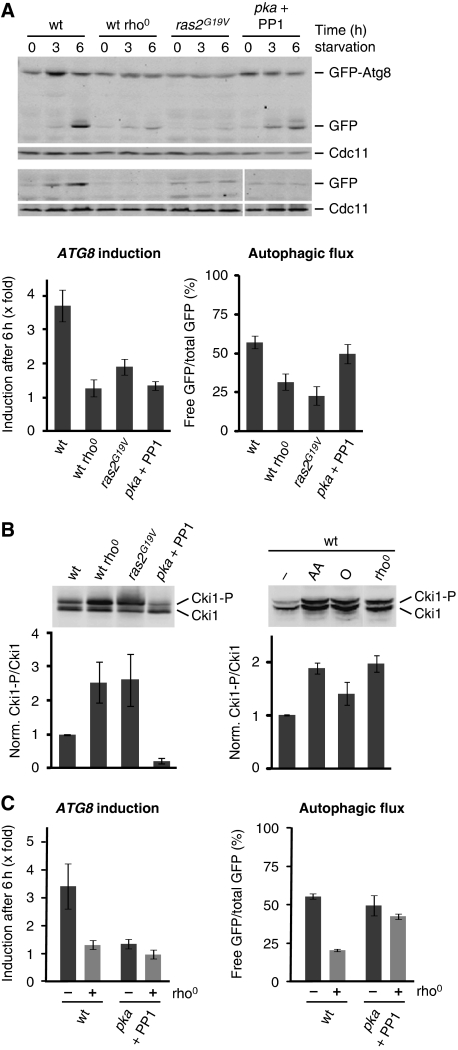
Mitochondrial function controls autophagy by modulating PKA activity. (A) PKA-dependent regulation of the autophagic response under amino-acid starvation. Wild-type, rho0, pka, and ras2G19V-expressing cells harbouring prATG8-GFP-ATG8 (upper panels) or prATG8-GFP (lower panels) were exposed to amino-acid starvation medium supplemented with galactose. PKA activity in pka was inhibited by addition of 1NM-PP1 (PP1; 1 μg/ml). Samples were analysed as described in Figure 1A; autophagic flux was determined after 3 and 6 h. (B) In vivo activity of PKA. Wild-type, rho0, pka, and ras2G19V-expressing cells harbouring 6xMYC-cki12−200(S125/130A) (Cki1) were grown in galactose medium. When indicated, wild-type cells were grown in galactose medium in the presence of antimycin A (AA) or oligomycin (O) for 6 h. PKA-dependent phosphorylation of Cki1 was analysed by whole cell extraction and western blot analysis using a α-Myc antibody (upper panels). Ratio of phosphorylated (Cki1-P) and non-phosphorylated (Cki1) forms of Cki1 relative to wild-type cells (wt=1) (lower panels). (C) PKA inhibition restores autophagic flux, but not ATG8 induction in the presence of mitochondrial dysfunction. Wild-type, rho0, pka, and pka rho0 cells expressing prATG8-GFP-ATG8 were treated as described in (A). Samples were analysed as described in Figure 1A. The means and s.d. of four (n=4) independent experiments are indicated in (A–C).

