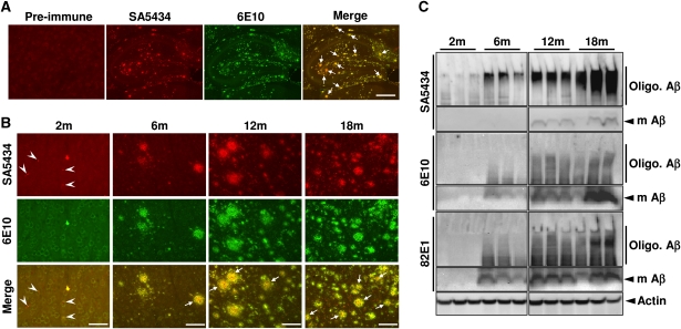
Figure 3.
Detection of pAβ in brains of APP transgenic mice. (A, B) Immunohistochemical detection of npAβ and pAβ by antibodies 6E10 and SA5434, respectively, in hippocampal regions of 9m mice (A) and in cortical regions of APPswe/PS1E9 double transgenic (tg) mouse brain of different ages (B). Plaques with pronounced reactivity of SA5434 in the core region are indicated by arrows. pAβ deposits selectively stained with SA5434 in 2m mice are indicated by arrowheads. The corresponding pre-immune sera (A) or SA5434 after pre-adsorption (Supplementary Figure S5) showed no specific staining. Scale bars represent 500 μm (A), 50 μm (B: 2m) and 200 μm (B: 6, 12 and 18m). (C) Biochemical analysis of npAβ and pAβ in mouse brain extracts. Brain homogenates of tg mice from 2 to 18 months (three mice for each age) were analysed by WB with antibodies SA5434, 6E10 and 82E1. Migrations of monomeric (m Aβ) and oligomeric Aβ (Oligo. Aβ) variants are indicated. The pronounced reactivity of SA5434 with smear in the upper part of the gels indicates the enrichment of pAβ in oligomeric assemblies. SA5434 did not detect pAβ in brain extracts of non-tg mice (Supplementary Figure S4).

