
Figure 4.
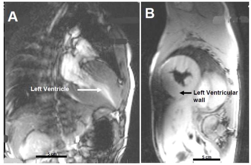
Cardiac MRI of a 10 year old male with Friedreich’s Ataxia. Panel A: Long axis view of hyperdynamic heart with contrast showing severe hypertrophy of left ventricular walls and mid-cavitary obliteration in systole. Ejection fraction = 81%. Panel B: Short axis of heart in systole showing hypertrophic cardiomyopathy. Bar = 5 cm in both panels.