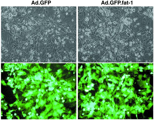
Figure 1.
Photomicrographs showing gene-transfer efficiency. Rat cardiac myocytes were infected with Ad.GFP (Left; control) or Ad.GFP.fat-1 (Right). Forty-eight hours after infection, cardiomyocytes were visualized with bright light (Upper) and at 510 nm of blue light (Lower). Coexpression of GFP demonstrates visually that the transgene is being expressed in cells in a high efficiency.

