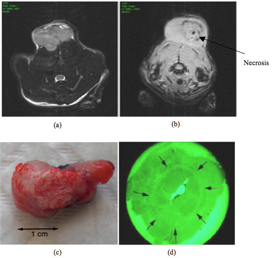
Figure 1.

(a) Pre-treatment MR image of the rat. (b) Post-treatment MR image with the visualization of the necrosis. The image is in a different plane than image (a). (c) The tumor after treatment and excision. (d) Histological tumor slice with green filter to enhance the circular limits of the coagolative necrosis (black arrows).
