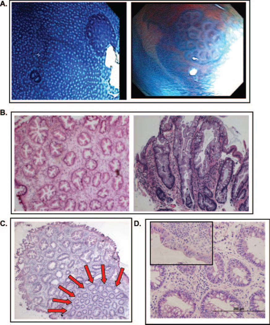
Fig. 1.
Macroscopic and histological analysis of ACF. (A) Gross views of ACF visualized through the Olympus prototype close-focus endoscope at a magnification of ×60. The colon epithelium was stained with 0.5% methylene blue. (B) H&E stained frozen sections of hyperplastic ACF, cross-sectional (left, ×200) and longitudinal (right, ×400) views. (C) H&E-stained frozen section of hyperplastic ACF with adjacent normal colonic mucosa indicated by the red arrows (×200). (D) H&E-stained frozen section of dysplastic ACF, cross-sectional view (×400). The inset in the upper left shows a region of surface dysplasia from the same lesion.

