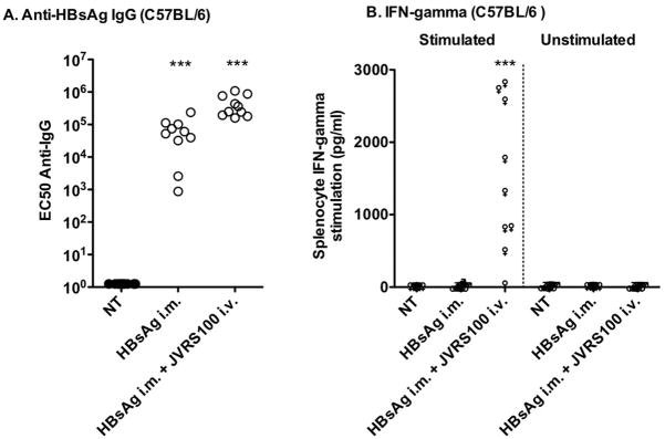
Figure 1.
Responses of A) HBsAg-specific IgG and B) IFN-γ to no treatment (NT), HBsAg (i.m., 5 μg), or HBsAg plus JVRS-100 (i.v., 10 μg) in female C57BL/6 mice (>6 weeks). Animals were treated on days 1, 22, and 43 and necropsied on day 57. Serum was assayed for HBsAg-specific IgG. The cell culture supernatants of splenocytes stimulated with HBsAg or unstimulated were assayed for IFN-γ by the same method shown in Figure 2. Ten animals were included in each group. JVRS-100 was made as follows (Morrey et al., 2008). A sterile 10 mM solution of cationic liposomes composed of DOTIM [octadecenoyloxy (ethyl-2-heptadecenyl-3-hydroxyethyl) imidazolinium chloride] and cholesterol was prepared in a 1:1 molar ratio as previously described (Dow et al., 1999; Gowen et al., 2006). A stock of 0.1 mg/mL was made by first dissolving the product in sterile water for injection to 1 mg/mL and then further diluting it into 5% Dextrose (Baxter, Deerfield Ill.) to a final dextrose concentration of 4.5%. Prior to injection, cationic liposomes were gently mixed with erroneous plasmid DNA (pMB75.6 empty vector lacking the downstream HCMV promoter) at a ratio of 16 nmol lipid per 1 μg DNA in 10% sucrose in water at room temperature. Ten micrograms of JVRS-100 was administered intravenously (i.v.) or intramuscularly (i.m.), respectively. Five micrograms of HBsAg (Biodesigns International, Maine) in sterile PBS was administered i.m. in 0.05 mL to each animal.***P ≤ 0.001 using one-way analysis of variance. Prism 4, GraphPad Software, Inc. was used for all statistical analyses.

