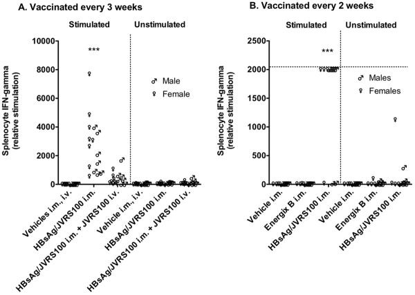
Figure 3.
IFN-γ responses to vehicle, JVRS-100 (IV, 10 μg), HBsAg (i.m., 5 μg), HBsAg and JVRS-100 (i.m., 5 μg, 10 μg, respectively) plus JVRS-100 (i.v., 10 μg), or HBsAg and JVRS-100 (i.m., 5 μg, 10 μg, respectively) in female and male HBV transgenic mice (>6 weeks, 23.6 ± 2.7 g). Animals were treated A) once every 3 weeks on days 1, 22, and 43 and necropsied on day 57, and B) once every 2 weeks on days 1, 14, 28, 42 and necropsied on day 57. Spleens were removed, minced and strained to isolate the splenocytes. For lysing the red blood cells, the splenocytes were incubated for 10 min at room temperature in lysing buffer. The solution was centrifuged, supernatant discarded and the packed cells were then washed twice in growth medium. Splenocyte concentration was then adjusted to 5 × 10⁶/mL. A volume of 2 mL of each sample was then stimulated with final concentration of 1ug/ml HBsAg for 48 hours in a CO2 incubator at 37°C. The cell culture supernatants of splenocytes were assayed for IFN-γ. IFN-γ secreted by splenocytes in response to HBsAg were measured (cat# 88-7314, Mouse ELISA Ready-SET-Go!, eBiosciences). This IFN-γ secretion would be primarily due to antigen-specific CD4 and CD8 T-cells generated following vaccination. Ten animals were included in each group. Horizontal line is the upper limit. (***P ≤ 0.001 compared with vehicle using one-way analysis of variance.)

