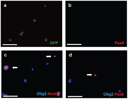
Figure 4. In vitro overexpression of Pax6 inhibits cell intrinsic expression of Olig2.
The olig2-GFP mouse SVZ was manually dissected from P4 pups. Tissue was washed in PBS, then incubated to 0.05%trypsin/0.53 mM EDTA (Invitrogen) at 37°C for 10 min and triturated. Trypsin was neutralized with PBS with 2%FBS. Neutralized cells were filtered and separated with 4%glucose PBS solution. Cell sorting was performed in the Columbia FACS facility using a modified triple laser FACS instrument and Summit software (DAKO) or DIVA software (BD Biosciences). The FlowJo program (Tree Star) was used for flow cytometry data analysis. Sorted Olig2-GFP cells were attached on glass slides and cultured in B1O4 conditioned medium (Hunter and Bottenstein, 1990; Hunter and Bottenstein, 1991) mixed 1∶3 with DMEM and then transfected with either pax6 or control eGFP expression constructs. Infected cells were kept in culture with basal media (DMEM, N2, T3, and penicillin/streptomycin/amphotericin) for 2d and then fixed in 4%paraformaldehyde, stained and analyzed by fluorescence microscopy. (a, b) After sorting, Olig2-eGFP positive cells were plated, incubated 7 days and immunostained with antibodies against Pax6. Virtually all of Olig2-eGFP+ cells (a) are Pax6-(b). eGFP immunostaining in green, Pax6 in red. (c) Sorted Olig2-eGFP cells were transfected with control Hcred vector. 48 hrs after transfection, immunostaining was done with anti-Olig2 antibody. Hcred immunostaining in red, Olig2 in Cy5. (d) Sorted Olig2-eGFP cells were transfected with the vector expressing flag-tagged wt Pax6. Each result was obtained by three independent experiments. Double immunostaining with anti-Olig2 and Pax6 antibodies demonstrates that transfected cells were Pax6+ but Olig2-. Olig2 immunostaining in Cy5, Pax6 in red. Scale bars, 50 µm.

