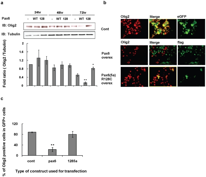
Figure 7. Overexpression of Pax6 decreases Olig2 protein levels.
(a) Proteins for Western blotting were analyzed 24, 48, and 72 hrs after transfection. (a) Oli-Neu cells were transfected with flag-tagged WT Pax6 (WT), Pax6(5a)R128C (128), or eGFP control vector (-). (b) Immunostaining analysis 72 hrs after transfection shows that overexpressed Pax6 decreases Olig2 levels in Oli-Neu cells. (c) Histogram summarizing percentage of Olig2+ cells in Oli-Neu cells after transfection. Each result represents a mean ± S.D. of three independent experiments. At each time point (24, 48, 72 h) we compared the Olig2 protein band seen after transfection with the WT Pax6 vector and the mutant Pax6 vector with the Olig2 band seen after transfection with the control vector. * by student's t test p<0.05, ** by student's t test p<0.01.

